How Exercise Can Cause Injury – NYC Sports Chiropractor Explains
The Relationship of Femoro Acetabular Impingement (FAI), Hip labral tears, Sports Hernia, and Athletic Pubalgia.
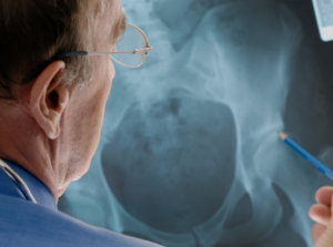
 In the last 20 years there has been a surge in the amount of Femoro Acetabular impingement (FAI) diagnosed. FAI is a condition where there is too much friction in the hip joint from bony irregularities causing pain and decreased range of hip motion. The femoral head and acetabulum rub against each other creating damage and pain to the hip joint. The damage can occur to the articular cartilage (the smooth surface of the ball or socket) or the labral tissue (the lining of the edge of the socket) during normal movement of the hip. The articular cartilage or labral tissue can fray or tear after repeated friction. In our midtown NYC sports chiropractic clinic, we most commonly see the early presentation of a chronic FAI in the 25-35 year old endurance athletes (marathon, triathletes, cyclists) or dancers/hockey/soccer players.
Is the way we exercise causing injury?
Researchers look for trends and contributing factors of athletes suffering this type of injury. Even though this condition is considered rare we see it more often in certain patient populations. The speed, intensity, and volume of training contributes to this condition becoming symptomatic. The early specialization in sports overloads some tissues and leads to poor development of others (e.g. hockey).
The increase in popularity of long distance sports contributes to an increase in prevalence of symptoms (e.g. couch to marathon programs) In the clinic we see this condition most commonly in someone that has an asymptomatic FAI and runs their first marathon or plays through a groin injury and the underlying FAI becomes symptomatic. If the cause is not addressed over time, the cartilage and labrum first frays or tears, then is lost until, eventually the femur bone and acetabulum bone impact on one other. Bone on bone friction is commonly referred to as Osteoarthritis.
Does our structure predispose us to this type of injury?
Normal anatomy of the hip is a ball and socket joint that distributes forces evenly as we flex, extend, and load the hip. You can thank your parents for the structure of your hip, and we can only play the hand we are given. If you get a pinch in the front of your hip when you flex and bring your hip across midline-it is a sign of some type of impingement. The shape of your hip can predispose an individual to impingement: you can have a CAM (more mass on the ball) and/or Pincer (extension off the socket) impingement, Coxa vara/valgus impingement due to the angle of the femoral neck, or Hip dysplasia (continuum diagnosis associated with a shallow hip socket and abnormal femoral neck angle are other conditions that lead to early labral degeneration.
FAI impingement generally occurs as two forms: Cam and Pincer.
CAM Impingement: when the femoral head and neck are not perfectly round, most commonly due to excess bone that has formed. This lack of roundness and excess bone causes abnormal contact between the surfaces.
PINCER Impingement: Is when the socket or acetabulum rim has overgrown and is too deep. It covers too much of the femoral head resulting in the labral cartilage being pinched. The Pincer form of impingement may also be caused when the hip socket is abnormally angled backwards causing abnormal impact between the femoral head and the rim of the acetabulum. Most diagnoses of FAI include a combination of the Cam and Pincer forms.
It’s a little bit of everything.
More athlete’s are suffering this type of injury than in the past due to early detection with advanced imaging techniques and the nature of how we exercise. Symptoms of FAI and labral tears found on imaging may resolve with conservative treatment, however if conservative treatment fails it may necessitate surgical repair. Sometimes symptoms persist in post-surgical repaired hips where the impingement was removed! We start to hear the terms “bursitis,” “capsulitis,” ”Tendonitis,” and to a lesser extent “sports hernia,” and “athletic pubalgia.” These terms have shown up in sports medicine circles and mainstream media coverage of sports injuries more frequently. At our clinic if the hip impingement was surgically removed it is our job to normalize the tone of tissue and remove any of the compensations. Many times it is the learned movement that causes the symptoms. The surgery takes away the impingement but doesn’t change the way you move.
There is no universally accepted definition of sports hernia.
With the increased popularity of these terms there is no universally accepted definition of sports hernia. It is sometimes used as shorthand for virtually any condition affecting the athlete’s lower abdomen and groin area. Furthermore there is a misnomer involved-in clinical examination there is no actual “hernia” found by medical definition(that classic bulge of tissue through muscle). A more appropriate term is athletic pubalgia, though it is less frequently heard.
Whatever you call it, this condition is a serious problem for Athletes.
The common ground is that sports hernia and athletic pubalgia is a combination of injuries affecting the groin and abdominal area making it more of a Syndrome than a specific injury. It is the result of chronic shearing forces across the pubic symphysis which functions as the pivot point for force transfer between the lumbopelvic and femoroacetabular joints. The chronic strain and shearing forces can lead to micro tears of the rectus abdominis muscle or its tendon at the pubic insertions. Movement of the femurs during athletic activity and activation of the adductors add to the stress generated by repetitive adductor muscle activation. Over time these forces cause progressive micro stress to the posterior abdominal wall, causing a separation of the transversalis fascia and internal oblique apneurosis from the inguinal ligament.
Chicken or the Egg?
Does the hip labrum fray and tear first then the altered function of the pelvis or vice a versa? Poor core stability can cause an excessive anterior pelvic tilt, increasing tension to the adductor compartment and femur. Poor adductor strength or flexibility will place increased stress on the pubic symphysis altering the lumbopelvic function and affecting various muscle attachments of the pelvis, including the obliques, rectus, transversalis muscles, and the conjoined tendon. Here at CSC+M, we work with physical therapy and ART (active release technique) 8 weeks pre-surgical and 8 weeks post-surgical to give conservative treatment the best chance of success.
Fix IT…Poor Core Stability, Poor Eccentric strength of the psoas, iliacus, and abdominals, Poor Eccentric strength of adductors.
Muscles heal via scar tissue that decreases tensile strength and elasticity. Treatment and rehabilitation of those injuries is important for maintaining dynamic stability between the muscles in the lumbopelvic system. Here at the clinic we utilize ART protocols on the adductor magnus & longus, pectineus, rectus abdominus, obliques, psoas, iliacus, quadratus lumborum and glutes. Our physical therapists focus on restoring lumbopelvic control, balancing the strength of the adductors (eccentric strength first), and single leg strength (building from the ground to half-kneeling position). Our Acupuncturist can target scar tissue with needles into muscles and into tendinous insertions. Cupping and massage can also be an effective way to release bound down scar tissue. We also use Graston protocols when necessary to restore myofascial integrity. Our medical staff uses rock tape and trigger point therapy to normalize movement along myofascial chains.
Commit to a conservative treatment plan to give your tissues the best chance recover and get back to active life.
